Home training device for the deep spinal muscles

Table of contents

1 Back pain affects many people

In response to my own back pain, I built a prototype intended to train the lumbar extensor muscles with the pelvis stabilized. The background to why I did that is explained below.
The background combines scientific literature, anatomy, mechanics and my personal experience. The scientific evidence and my individual observations are identified separately below.
The journey to building the device began for me with back pain in the lumbar region. I was in my mid-30s, had trained for about 15 years, had never been overweight or smoked, and drank little alcohol. Those factors can reduce some health risks, but they do not rule out low back pain. So why do I have back pain?

1.1 Back pain is a major health burden

It seems as if everyone knows someone who has back pain. A quick search in the literature explains the reasons. Low back pain (LBP) is a leading cause of disability worldwide [1] [21]. Historical estimates illustrate its economic burden: a 1991 US review estimated annual costs of at least $50 billion [2], while a study using 2005 Swiss data estimated costs of 1.6% of GDP [3]. These figures should not be read as current cost estimates, but the global health burden remains substantial. Why can't we get back pain under control?

1.2 Understanding the causes of back pain

In most cases, LBP cannot be attributed to one clearly identifiable structure and is described as non-specific or chronic primary low back pain [21] [22]. Its course can be influenced by interacting biological, psychological, social, lifestyle and occupational factors. Age, smoking, high body weight and physical workload are associated with risk at population level [4] [5] [6] [7], but none determines whether an individual will have pain.
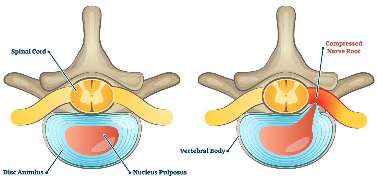
The complexity is also visible in spinal imaging. A disc herniation occurs when disc material is displaced through a weakened or torn annulus. It may cause pain when nerve irritation, inflammation or mechanical compression is clinically relevant, but disc abnormalities are also common in people without symptoms [8]. Imaging findings therefore need to be interpreted together with a person's symptoms and examination.

Spinal Anatomy Diagram

Figure 1: Herniated disc.

2 A special muscle

In the next few sections, I would like to give the reader the basic knowledge needed to clearly identify the problem of back pain. I will concentrate on the essentials in order to keep the format of this text as compact as possible.

2.1 The location of the multifidus

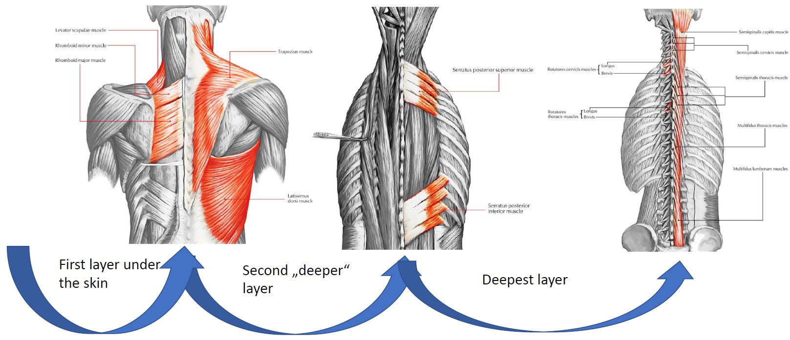
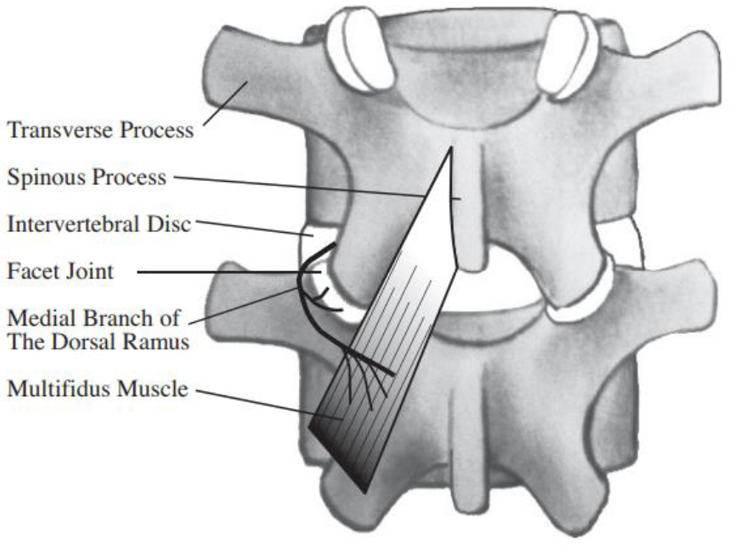
Let's imagine we are standing behind a person and looking at their back. Under the skin there is a layer of muscle called the "superficial muscle group". Muscles that lie under this layer are called the "intermediate muscle group". Another layer "deeper" is the "deep muscle group". Having reached the "deepest" level, we can now see our target. The (lumbar) spine and some smaller muscle groups that are directly connected to the spine. There we find the so-called "lumbar extensor muscles". The so-called multifidus is one important part of this muscle group. Its fascicles run along the spine and span several vertebral segments (Fig. 3).

Spinal Anatomy Diagram

Figure 2: Different layers of the back muscles.

Spinal Anatomy Diagram

Figure 3: Multifidus muscle.

2.2 The function of the multifidus

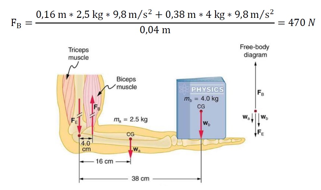
Muscles produce force by contracting. Depending on the task, that force can create movement, resist movement or stabilize a joint. A short physics problem will enable us to better estimate the forces that muscles normally have to exert in order to be able to carry out our everyday tasks. Let's assume someone is lifting a 4 kg object in their hand (Fig. 4). Now we want to calculate how much force the biceps has to exert.

Spinal Anatomy Diagram

Spinal Anatomy Diagram

Figure 4: Lever law on the human body.

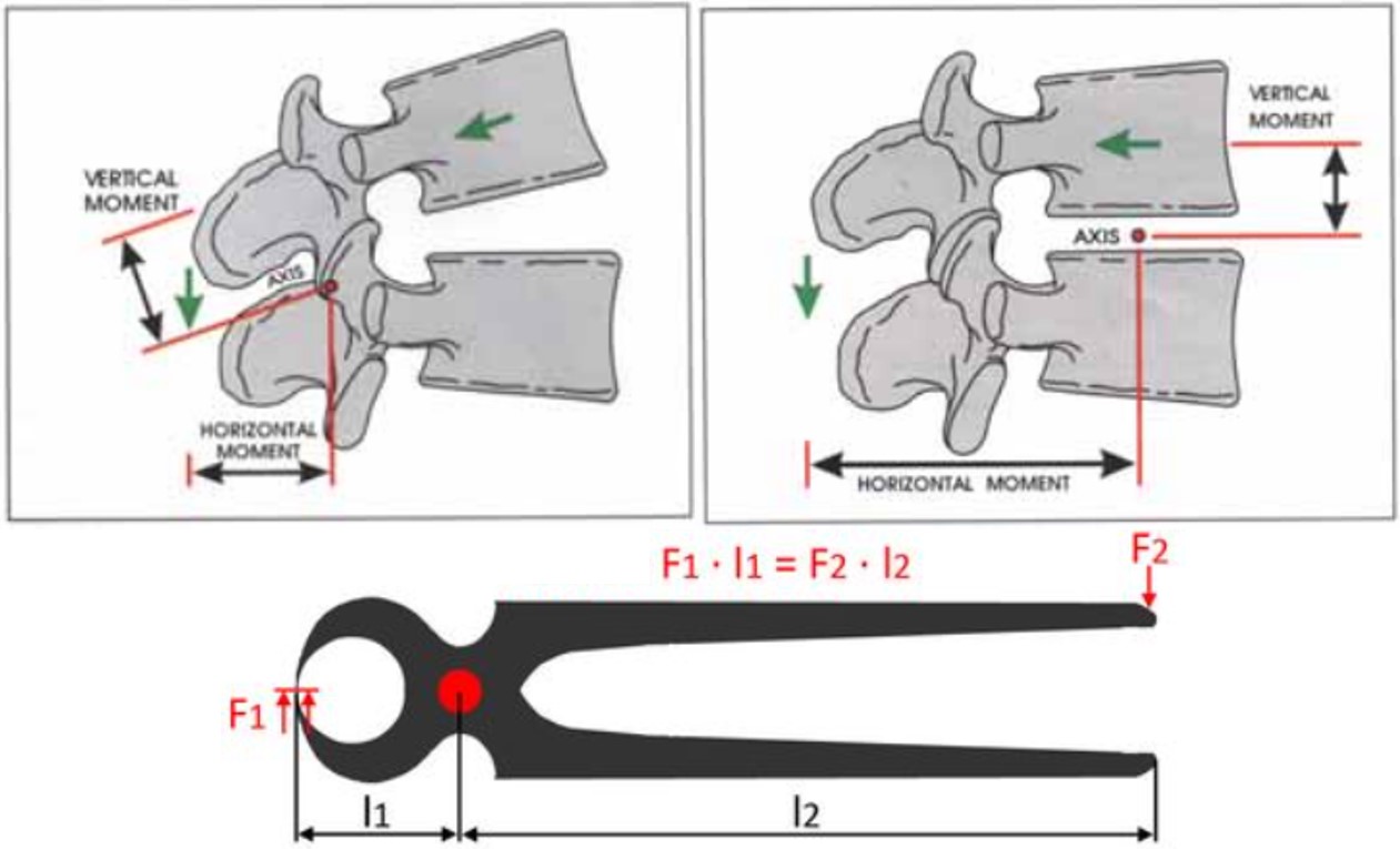
In this simplified example, the arm and the external weight exert approximately 64 N downward, while the calculated biceps force is about 470 N because the muscle acts through a short moment arm. The spine is mechanically more complex than this single-joint model: several muscles and passive tissues share loads, and measured external torque cannot be converted directly into force produced by the multifidus alone. Figure 5 illustrates a lever concept, not a validated estimate of multifidus force.

Spinal Anatomy Diagram

Figure 5: Simplified lever concept related to spinal motion.

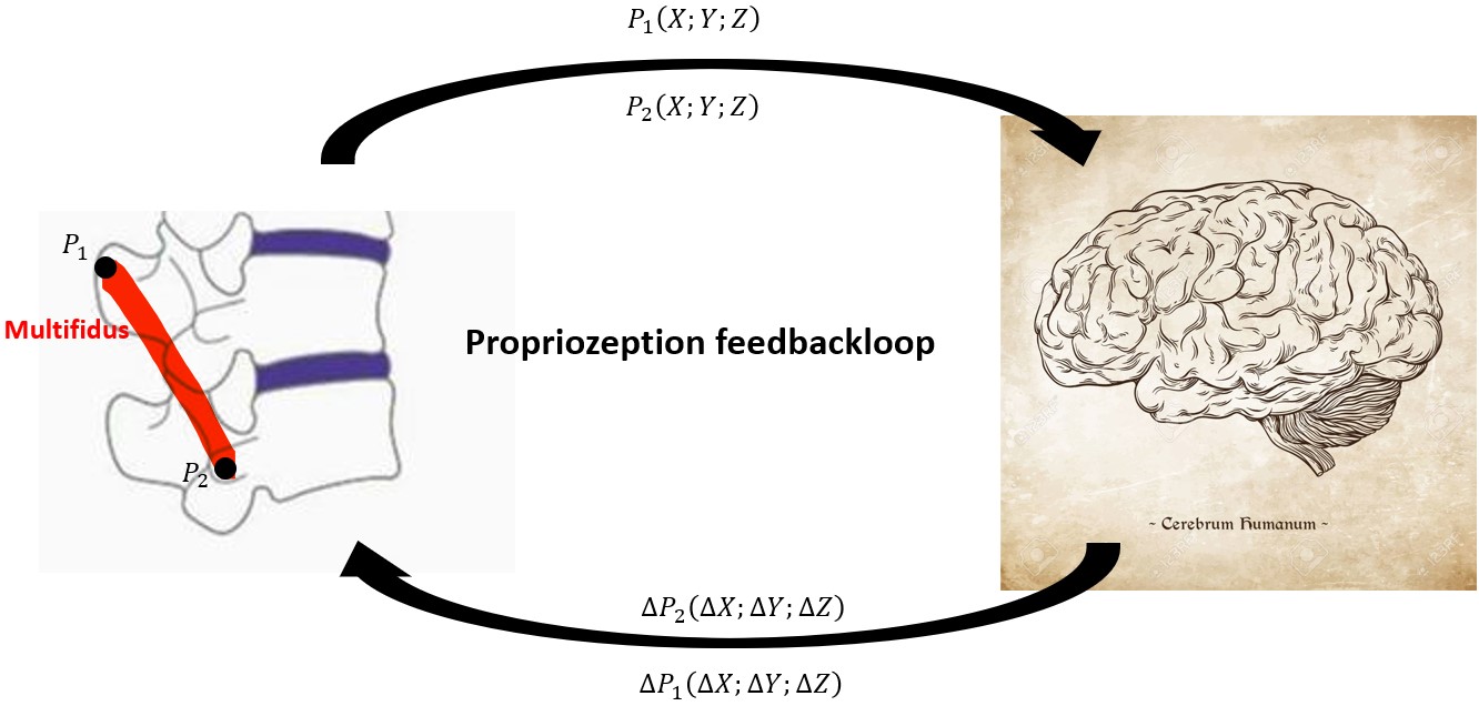
Electromyography studies show that multifidus activation varies with posture, load, movement direction and the depth and spinal level being measured [9] [10]. It can be active during standing, walking, lifting, flexion, extension and rotation. It contributes to lumbar extension and rotation, controls movement between vertebral segments and contains many sensory receptors that support proprioception. Proprioception is the nervous system's awareness of body position and movement; it is one function of the multifidus, not the muscle's only function (Fig. 6).

Spinal Anatomy Diagram

Figure 6: Proprioception principle at the multifidus muscle.

The multifidus is therefore an important contributor to spinal movement and control. Dysfunction may be relevant in some people with LBP, but it is not established as the single centre or cause of back pain.

2.3 Multifidus and back pain

Studies frequently report differences in multifidus morphology between groups with and without LBP. The most commonly studied features are reduced cross-sectional area, asymmetry and myosteatosis, meaning increased fat within and between muscle tissue. Reduced size does not automatically mean reduced strength, and these changes can have several causes, including pain-related inhibition, reduced activity, ageing, nerve injury, metabolic factors and spinal pathology.
Most studies in this area are observational. They can show an association, but usually cannot determine whether a muscle change preceded the pain, resulted from it, or is influenced by a third factor [23]. Examples include:

  • Study[11] examined multifidus muscle size in 80 people (15-18 years old).
    Result: Adolescents with LBP had smaller multifidus measurements on ultrasound. The study does not establish that smaller muscles caused the pain.
  • Study[12] examined multifidus muscles in 78 people (17-72 years old).
    Result: Multifidus atrophy was observed in 80% of this clinical LBP sample and was associated with leg pain. The authors noted that cause and effect were unknown.
  • Study[13] examined multifidus muscles in 90 people.
    Result: Group differences in multifidus size and symmetry were observed, particularly at lower lumbar levels.
  • Study[14] examined multifidus muscles in 132 people with a herniated disc.
    Result: Compared with 132 controls, the disc-herniation group showed differences in multifidus size and fatty infiltration. This retrospective study demonstrates correlation, not causation.
  • Study[15] examined multifidus muscles in 23 professional football players with LBP.
    Result: Players with LBP showed differences in trunk muscle size or function and hip strength. The sample was small and sport-specific.

Overall conclusion: people with LBP have, on average, somewhat smaller multifidus muscles and more intramuscular fat than pain-free controls [23]. This supports studying the muscle as one potentially relevant factor. It does not show that multifidus dysfunction is present in everyone with LBP or that strengthening it will prevent or resolve every case. Exercise is recommended for many people with persistent LBP, with the programme matched to the individual rather than focused on one muscle by default [21] [24].

3 Lumbar-extension training with pelvic stabilization

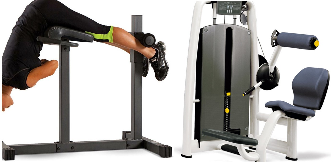
For the lower back area, fitness equipment such as that shown in Figure 7 is usually available.

Spinal Anatomy Diagram

Figure 7: Typical training equipment for training the lower back.

These exercises can activate the lumbar extensor muscles and improve outcomes such as endurance. However, movement of the pelvis allows the gluteal and hamstring muscles to contribute, so the exercise is less specific to lumbar extension. Pelvic stabilization was developed to reduce that contribution and make lumbar-extensor testing and training more isolated.

3.1 MedX lumbar extension therapy device

In 1986, Arthur Jones founded the company MedX in the USA. In collaboration with the University of Florida, after years of development, they released medical training and measuring devices. The MedX LE Lumbar Extension is one device used to measure and train lumbar-extension torque. Its development is described in the book "The Lumbar Spine, The Cervical Spine And the Knee; Testing and Rehabilitation". Research using this type of device supports an important mechanical point:
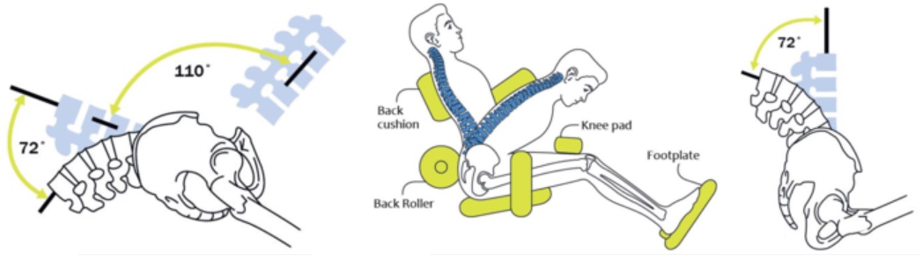
Stabilizing the pelvis reduces pelvic rotation and contribution from the hip extensors, allowing lumbar-extension torque to be tested and trained more specifically [20]. The exercise still involves a group of lumbar extensor muscles; it does not isolate or measure the multifidus alone. The stabilization also reduces the available movement from combined trunk and hip motion to a smaller lumbar range, commonly about 72 degrees on this device.

Spinal Anatomy Diagram

Figure 8: MedX lumbar extrension schematics.

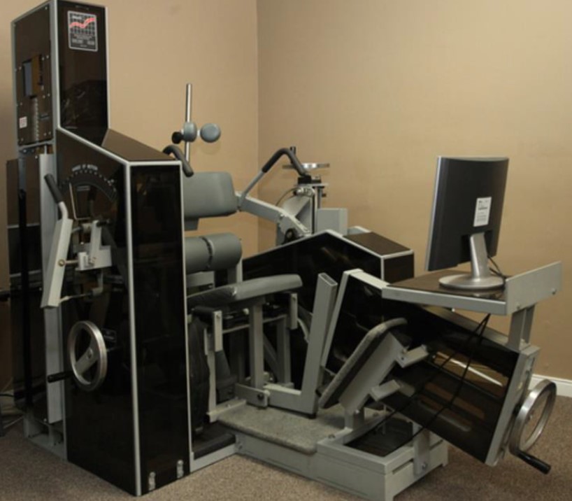
The MedX LE device is shown in Figure 9.

Spinal Anatomy Diagram

Figure 9: Original MedX lumbar extension.

Isolated lumbar-extension exercise has been studied as one option for people with LBP. Individual trials report improvements, but their designs and populations differ, and the certainty of the overall evidence is very low. A recent systematic review found a possible reduction in pain, with less consistent effects on disability and physical function [25].

Taken together, the evidence supports pelvic stabilization when the goal is to isolate lumbar-extension torque. It also suggests that isolated lumbar-extension exercise may help some people with LBP, but it has not been established as superior to all other exercise approaches or as a universal treatment [24] [25].
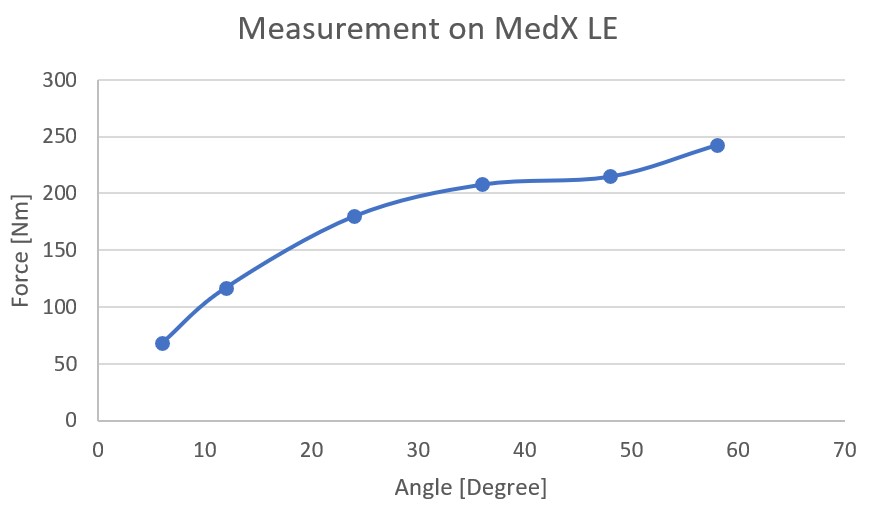
It turned out that a therapist about 100 km away from me had such a device. So I went there to have my back muscles measured. The measurement result is shown in Figure 10.

Spinal Anatomy Diagram

Figure 10: Measurements with a MedX LE.

My measured torque was below the device's comparison curve. This showed lower performance on that particular test, but it could not diagnose the cause of my pain or identify the strength of the multifidus alone. I nevertheless decided to explore this training approach. Every time I wanted to train on the "original device" I would have to drive 200 km and pay 50 euros. So I considered buying a home version of the machine. At the time, I could not find a compact home device that matched the stabilization and loading concept I wanted.

So I decided to build an experimental prototype.

3.2 Experimental home lumbar-extension device

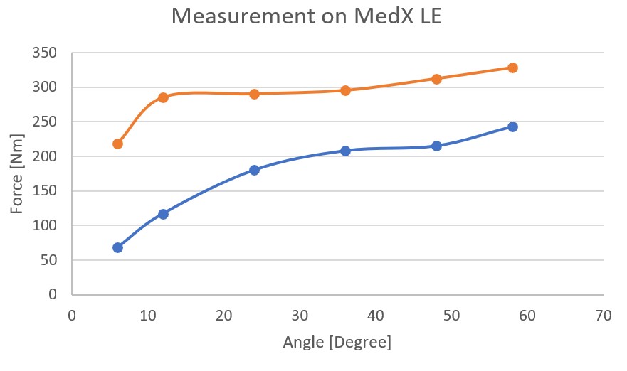
After a few months of tinkering, I thought I had built a usable prototype for training my lumbar extensor muscles with the pelvis stabilized. I trained with it once a week for 12 weeks and then went back to the MedX LE to take measurements. The result is the orange line in Figure 11.

Spinal Anatomy Diagram

Figure 11: Measurements with a MedX LE after training with the prototype.

The second test showed higher lumbar-extension torque at every measured angle, with an increase of more than 100% at the most extended position. My back pain also decreased during this period. This is an individual before-and-after observation, not a clinical trial: there was no control condition, blinding or repeated baseline, and the result may include training, familiarization with the test, effort, setup differences and natural variation. It does not establish that the prototype caused the pain reduction or that the gain came specifically from the multifidus. The graph reports torque in newton-metres (Nm), not muscle force.
Before I describe my device in more detail, I would like to briefly mention my original train of thought:

Features I considered unnecessary for a basic training prototype, compared with the MedX LE, include:

Design features intended to approximate controlled lumbar-extension exercise:

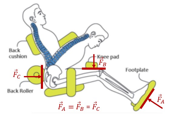
The tensioning device must in principle act in at least three places (Fig. 12, points A, B, C). Tension is applied in one place and this force must be counteracted in two places.

Spinal Anatomy Diagram

Figure 12: Working principle for a home training device.

The pivoting cushion (point C) provides a visible indication of pelvic movement. If it rotates during the exercise, pelvic motion is occurring and the setup may not be providing the intended stabilization. A stationary cushion does not prove that the multifidus is isolated or quantify which muscles are producing the torque; validating that would require biomechanical and physiological measurements.
Under prototype one can find out more about the current version.

Safety notice: This prototype is experimental and has not been clinically validated or certified as a medical device. Loaded spinal exercise is not appropriate for everyone. Anyone with back pain should obtain appropriate medical or physiotherapy advice before using an experimental device. New bowel or bladder problems, saddle numbness, progressive leg weakness, fever, unexplained weight loss, major trauma or a history of cancer require prompt medical assessment [27].

4 Bibliography

[1] GBD 2015 Disease and Injury Incidence and Prevalence Collaborators, “Global, regional, and national incidence, prevalence, and years lived with disability for 310 diseases and injuries, 1990-2015: a systematic analysis for the Global Burden of Disease Study 2015,” Lancet Lond. Engl., vol. 388, no. 10053, pp. 1545–1602, Oct. 2016, doi: 10.1016/S0140-6736(16)31678-6.
[2] J. W. Frymoyer and W. L. Cats-Baril, “An overview of the incidences and costs of low back pain,” Orthop. Clin. North Am., vol. 22, no. 2, pp. 263–271, Apr. 1991.
[3] S. Wieser et al., “Cost of low back pain in Switzerland in 2005,” Eur. J. Health Econ. HEPAC Health Econ. Prev. Care, vol. 12, no. 5, pp. 455–467, Oct. 2011, doi: 10.1007/s10198-010-0258-y.
[4] P. Kjaer, N. Wedderkopp, L. Korsholm, and C. Leboeuf-Yde, “Prevalence and tracking of back pain from childhood to adolescence,” BMC Musculoskelet. Disord., vol. 12, p. 98, May 2011, doi: 10.1186/1471-2474-12-98.
[5] C. E. Dionne, K. M. Dunn, and P. R. Croft, “Does back pain prevalence really decrease with increasing age? A systematic review,” Age Ageing, vol. 35, no. 3, pp. 229–234, May 2006, doi: 10.1093/ageing/afj055.
[6] R. Shiri, J. Karppinen, P. Leino-Arjas, S. Solovieva, and E. Viikari-Juntura, “The association between smoking and low back pain: a meta-analysis,” Am. J. Med., vol. 123, no. 1, p. 87.e7–35, Jan. 2010, doi: 10.1016/j.amjmed.2009.05.028.
[7] T.-T. Zhang, Z. Liu, Y.-L. Liu, J.-J. Zhao, D.-W. Liu, and Q.-B. Tian, “Obesity as a Risk Factor for Low Back Pain: A Meta-Analysis,” Clin. Spine Surg., vol. 31, no. 1, pp. 22–27, Feb. 2018, doi: 10.1097/BSD.0000000000000468.
[8] S. D. Boden, D. O. Davis, T. S. Dina, N. J. Patronas, and S. W. Wiesel, “Abnormal magnetic-resonance scans of the lumbar spine in asymptomatic subjects. A prospective investigation,” J. Bone Joint Surg. Am., vol. 72, no. 3, pp. 403–408, Mar. 1990.
[9] J. P. Arokoski et al., “Back and hip extensor muscle function during therapeutic exercises,” Arch. Phys. Med. Rehabil., vol. 80, no. 7, pp. 842–850, Jul. 1999, doi: 10.1016/s0003-9993(99)90237-x.
[10] F. P. Valencia and R. R. Munro, “An electromyographic study of the lumbar multifidus in man,” Electromyogr. Clin. Neurophysiol., vol. 25, no. 4, pp. 205–221, Jun. 1985.
[11] N. Rahmani, A. Kiani, M. A. Mohseni-Bandpei, and I. Abdollahi, “Multifidus muscle size in adolescents with and without back pain using ultrasonography,” J. Bodyw. Mov. Ther., vol. 22, no. 1, pp. 147–151, Jan. 2018, doi: 10.1016/j.jbmt.2017.05.016.
[12] D. F. Kader, D. Wardlaw, and F. W. Smith, “Correlation between the MRI changes in the lumbar multifidus muscles and leg pain,” Clin. Radiol., vol. 55, no. 2, pp. 145–149, Feb. 2000, doi: 10.1053/crad.1999.0340.
[13] J. Hides, C. Gilmore, W. Stanton, and E. Bohlscheid, “Multifidus size and symmetry among chronic LBP and healthy asymptomatic subjects,” Man. Ther., vol. 13, no. 1, pp. 43–49, Feb. 2008, doi: 10.1016/j.math.2006.07.017.
[14] C. Liu et al., “Is there a correlation between upper lumbar disc herniation and multifidus muscle degeneration? A retrospective study of MRI morphology,” BMC Musculoskelet. Disord., vol. 22, no. 1, p. 92, Jan. 2021, doi: 10.1186/s12891-021-03970-x.
[15] J. A. Hides, T. Oostenbroek, M. M. Franettovich Smith, and M. D. Mendis, “The effect of low back pain on trunk muscle size/function and hip strength in elite football (soccer) players,” J. Sports Sci., vol. 34, no. 24, pp. 2303–2311, Dec. 2016, doi: 10.1080/02640414.2016.1221526.
[16] B. W. Nelson, E. O’Reilly, M. Miller, M. Hogan, J. A. Wegner, and C. Kelly, “The clinical effects of intensive, specific exercise on chronic low back pain: a controlled study of 895 consecutive patients with 1-year follow up,” Orthopedics, vol. 18, no. 10, pp. 971–981, Oct. 1995, doi: 10.3928/0147-7447-19951001-05.
[17] J. Steele, S. Bruce-Low, D. Smith, D. Jessop, and N. Osborne, “A randomized controlled trial of limited range of motion lumbar extension exercise in chronic low back pain,” Spine, vol. 38, no. 15, pp. 1245–1252, Jul. 2013, doi: 10.1097/BRS.0b013e318291b526.
[18] S. Ju, G. Park, and E. Kim, “Effects of an Exercise Treatment Program on Lumbar Extensor Muscle Strength and Pain of Rehabilitation Patients Recovering from Lumbar Disc Herniation Surgery,” J. Phys. Ther. Sci., vol. 24, no. 6, pp. 515–518, 2012, doi: 10.1589/jpts.24.515.
[19] J. L. Verna, J. M. Mayer, V. Mooney, E. A. Pierra, V. L. Robertson, and J. E. Graves, “Back extension endurance and strength: the effect of variable-angle roman chair exercise training,” Spine, vol. 27, no. 16, pp. 1772–1777, Aug. 2002, doi: 10.1097/00007632-200208150-00016.
[20] J. E. Graves et al., “Pelvic stabilization during resistance training: its effect on the development of lumbar extension strength,” Arch. Phys. Med. Rehabil., vol. 75, no. 2, pp. 210–215, Feb. 1994.
[21] World Health Organization, WHO guideline for non-surgical management of chronic primary low back pain in adults in primary and community care settings, Geneva, 2023, ISBN 978-92-4-008178-9.
[22] J. Hartvigsen et al., “What low back pain is and why we need to pay attention,” Lancet, vol. 391, no. 10137, pp. 2356–2367, 2018, doi: 10.1016/S0140-6736(18)30480-X.
[23] T. Seyedhoseinpoor et al., “Alteration of lumbar muscle morphology and composition in relation to low back pain: a systematic review and meta-analysis,” Spine J., vol. 22, no. 4, pp. 660–676, 2022, doi: 10.1016/j.spinee.2021.10.018.
[24] J. A. Hayden, J. Ellis, R. Ogilvie, A. Malmivaara, and M. W. van Tulder, “Exercise therapy for chronic low back pain,” Cochrane Database Syst. Rev., no. 9, CD009790, 2021, doi: 10.1002/14651858.CD009790.pub2.
[25] R. Trybulski et al., “Impact of isolated lumbar extension strength training on reducing nonspecific low back pain, disability, and improving function: a systematic review and meta-analysis,” Sci. Rep., vol. 15, 2025, doi: 10.1038/s41598-025-90699-5.
[26] T. Oosterhuis et al., “Rehabilitation after lumbar disc surgery,” Cochrane Database Syst. Rev., no. 3, CD003007, 2014, doi: 10.1002/14651858.CD003007.pub3.
[27] National Institute for Health and Care Excellence, Low back pain and sciatica in over 16s: assessment and management, NG59, updated 2020.